Figure 2
Atypical presentation of congenital pneumonia: Value of lung ultrasound
Virginie Meau-Petit* and Grenville F Fox
Published: 29 March, 2021 | Volume 4 - Issue 1 | Pages: 033-034

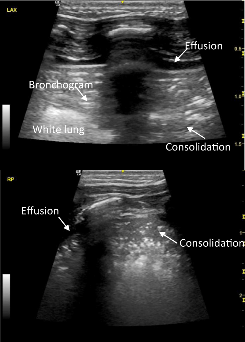
Figure 2:
Lung ultrasound images: left posterior axillary (a) and right posterior (b) views, showing small effusion facing large consolidation with bronchogram and irregular borders.
Read Full Article HTML DOI: 10.29328/journal.japch.1001027 Cite this Article Read Full Article PDF
More Images
Similar Articles
-
Glycemic status and its effect in Neonatal Sepsis - A prospective study in a Tertiary Care Hospital in NepalBadri Kumar Gupta*,Binod Kumar Gupta,Amit Kumar Shrivastava,Pradeep Chetri. Glycemic status and its effect in Neonatal Sepsis - A prospective study in a Tertiary Care Hospital in Nepal. . 2019 doi: 10.29328/journal.japch.1001006; 2: 015-019
-
Atypical presentation of congenital pneumonia: Value of lung ultrasoundVirginie Meau-Petit*,Grenville F Fox. Atypical presentation of congenital pneumonia: Value of lung ultrasound. . 2021 doi: 10.29328/journal.japch.1001027; 4: 033-034
-
Rationality and irrationality in the use of antibiotics in the epiclatino Latin American Neonatal UnitsAngela B Hoyos*. Rationality and irrationality in the use of antibiotics in the epiclatino Latin American Neonatal Units. . 2021 doi: 10.29328/journal.japch.1001028; 4: 035-037
-
Efficacy of intravenous immunoglobulins in the prophylaxis of neonatal sepsisSharif S*,Bloomer C,Al Assaf N,Khan R. Efficacy of intravenous immunoglobulins in the prophylaxis of neonatal sepsis. . 2021 doi: 10.29328/journal.japch.1001029; 4: 038-041
-
Clinical picture of pulmonary plague observed in the paediatric wards of antananarivoRivo Lova Herilanto Rakotomalala*,Lovasoa Randriamanga,Mihary Soa Karol,Mioramalala Sedera Aurélien,Annick Lalaina Robinson. Clinical picture of pulmonary plague observed in the paediatric wards of antananarivo. . 2021 doi: 10.29328/journal.japch.1001031; 4: 046-049
-
Rapidly involuting congenital hemangioma associated with Kasabach-Merritt SyndromeRosalia Ballona*,Jean Zevallos,Jeanette Núñez. Rapidly involuting congenital hemangioma associated with Kasabach-Merritt Syndrome. . 2021 doi: 10.29328/journal.japch.1001032; 4: 050-052
-
Revisiting childhood pneumonia in low-recourse setting hospitalsKarimeldin MA Salih*. Revisiting childhood pneumonia in low-recourse setting hospitals. . 2021 doi: 10.29328/journal.japch.1001035; 4: 062-066
-
The role of urine metabolomics among newborn infants with hypoxic ishaemic encephalopathy: a literature reviewNese Gadzama*,Irfan Ahmed,Sundus Khursheed,Niazy AL-Assaf,Rizwan Khan. The role of urine metabolomics among newborn infants with hypoxic ishaemic encephalopathy: a literature review. . 2021 doi: 10.29328/journal.japch.1001042; 4: 109-113
-
Severe brimonidine eye drop intoxication in a neonate as an accidental oral ingestionMusa Silahli*. Severe brimonidine eye drop intoxication in a neonate as an accidental oral ingestion. . 2022 doi: 10.29328/journal.japch.1001044; 5: 001-003
-
Effectiveness of different cord care interventions on omphalitis prevention among neonates in Nakuru County Hospital, KenyaLucy Kinanu*,James Mwaura,Anne Karani,Margaret Chege. Effectiveness of different cord care interventions on omphalitis prevention among neonates in Nakuru County Hospital, Kenya. . 2022 doi: 10.29328/journal.japch.1001046; 5: 015-021
Recently Viewed
-
Maternal and perinatal outcomes of uterine rupture in Lubumbashi, Democratic Republic of CongoJacques Ngoy Kitenge,Olivier Mukuku*,Xavier K Kinenkinda,Prosper L Kakudji. Maternal and perinatal outcomes of uterine rupture in Lubumbashi, Democratic Republic of Congo. Clin J Obstet Gynecol. 2020: doi: 10.29328/journal.cjog.1001067; 3: 136-141
-
Male in Early Adolescence Presenting with Guillain-Barré Syndrome Following BECOV2D VaccineVinit Suri*, Kanika Suri, Kunal Suri, Priyal. Male in Early Adolescence Presenting with Guillain-Barré Syndrome Following BECOV2D Vaccine. J Child Adult Vaccines Immunol. 2023: doi: 10.29328/journal.jcavi.1001010; 7: 001-004
-
Case Report of a Child with Beta Thalassemia Major in a Tribal Region of IndiaNeha Chauhan, Prakash Narayan, Mahesh Narayan, Manisha Shukla*. Case Report of a Child with Beta Thalassemia Major in a Tribal Region of India. J Child Adult Vaccines Immunol. 2023: doi: 10.29328/journal.jcavi.1001011; 7: 005-007
-
Health management using probioticsNeha Bisht,Amar P Garg*. Health management using probiotics. J Adv Pediatr Child Health. 2023: doi: 10.29328/journal.japch.1001053; 6: 001-006
-
Hereditary spherocytosis: review of cases and discussion of hematologic characteristics and updated diagnostic testingKennedy A Sabharwal,Michael W Simon*. Hereditary spherocytosis: review of cases and discussion of hematologic characteristics and updated diagnostic testing. J Adv Pediatr Child Health. 2023: doi: 10.29328/journal.japch.1001054; 6: 007-013
Most Viewed
-
Feasibility study of magnetic sensing for detecting single-neuron action potentialsDenis Tonini,Kai Wu,Renata Saha,Jian-Ping Wang*. Feasibility study of magnetic sensing for detecting single-neuron action potentials. Ann Biomed Sci Eng. 2022 doi: 10.29328/journal.abse.1001018; 6: 019-029
-
Evaluation of In vitro and Ex vivo Models for Studying the Effectiveness of Vaginal Drug Systems in Controlling Microbe Infections: A Systematic ReviewMohammad Hossein Karami*, Majid Abdouss*, Mandana Karami. Evaluation of In vitro and Ex vivo Models for Studying the Effectiveness of Vaginal Drug Systems in Controlling Microbe Infections: A Systematic Review. Clin J Obstet Gynecol. 2023 doi: 10.29328/journal.cjog.1001151; 6: 201-215
-
Causal Link between Human Blood Metabolites and Asthma: An Investigation Using Mendelian RandomizationYong-Qing Zhu, Xiao-Yan Meng, Jing-Hua Yang*. Causal Link between Human Blood Metabolites and Asthma: An Investigation Using Mendelian Randomization. Arch Asthma Allergy Immunol. 2023 doi: 10.29328/journal.aaai.1001032; 7: 012-022
-
Impact of Latex Sensitization on Asthma and Rhinitis Progression: A Study at Abidjan-Cocody University Hospital - Côte d’Ivoire (Progression of Asthma and Rhinitis related to Latex Sensitization)Dasse Sery Romuald*, KL Siransy, N Koffi, RO Yeboah, EK Nguessan, HA Adou, VP Goran-Kouacou, AU Assi, JY Seri, S Moussa, D Oura, CL Memel, H Koya, E Atoukoula. Impact of Latex Sensitization on Asthma and Rhinitis Progression: A Study at Abidjan-Cocody University Hospital - Côte d’Ivoire (Progression of Asthma and Rhinitis related to Latex Sensitization). Arch Asthma Allergy Immunol. 2024 doi: 10.29328/journal.aaai.1001035; 8: 007-012
-
An algorithm to safely manage oral food challenge in an office-based setting for children with multiple food allergiesNathalie Cottel,Aïcha Dieme,Véronique Orcel,Yannick Chantran,Mélisande Bourgoin-Heck,Jocelyne Just. An algorithm to safely manage oral food challenge in an office-based setting for children with multiple food allergies. Arch Asthma Allergy Immunol. 2021 doi: 10.29328/journal.aaai.1001027; 5: 030-037

If you are already a member of our network and need to keep track of any developments regarding a question you have already submitted, click "take me to my Query."


